|
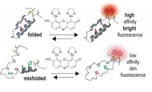
Schematic of fluorescent detector: When a target
protein is folded correctly, "tags" come together so that the dye
binds with high affinity and fluoresces brightly; misfolded
proteins have low affinity for the dye.
Credit: Schepartz/Nature Chemical Biology
|
�Our approach bypasses many of the problems
associated with fluorescent proteins, so that we can image protein
interactions in living cells,� said senior author Alanna Schepartz,
the Milton Harris Professor of Chemistry, and Howard Hughes Medical
Institute Professor at Yale. �Using these molecules we can
differentiate alternative or misfolded proteins from those that are
folded correctly and also detect protein partnerships in live cells.�
Each protein is a three-dimensional structure created by �folding� its
linear chain of amino acids. Usually only one shape �works� for each
protein. The particular shape a protein takes depends on its amino
acids and on other processes within the cell.
Schepartz and her team devised their new tagging system using small
molecules, called �profluorescent� biarsenal dyes. These molecules
easily enter cells and become fluorescent when they bind to a specific
amino acid tag sequence within a protein. While these compounds have
been used for about a decade to bind single proteins, this is the
first time they have been used to identify interactions between
proteins.
The researchers� strategy was to split the amino acid tag for the dye
into two pieces, locating each piece of the tag far apart in the chain
of a protein they genetically engineered and expressed in the cells.
Then they monitored cells exposed to the dye. Where the protein folded
correctly, the two parts of the tag came together and the fluorescent
compound bound and lit up. There was no signal unless the protein
folded normally.
�This method of detection can provide important insights into how
proteins choose their partners within the cell -
choices that may be very different from those made in a test tube,�
said Schepartz. She emphasizes that this technology does not monitor
the process of protein folding - but, rather �sees� the protein
conformations that exist at a given time.
�In theory, our technique could be used to target and selectively
inactivate specific protein complexes in the cell, as therapy, or to
visualize conformations at very high resolution for diagnostic
purposes,� said Schepartz. She speculates that the technology could be
applied to detection strategies that identify protein misfolding in
neurodegenerative diseases like Alzheimer�s or Parkinson�s.
|