|
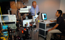
Philip Low, Purdue's Ralph C. Corley Distinguished
Professor of Chemistry, discusses a new cancer detection method
with graduate student Wei He (seated). Low's research team is able
to detect and count circulating tumor cells by shining a laser on
surface veins. The team uses a two-photon fluorescence microscope
(shown) to detect tumor cells labeled with tumor-specific
fluorescent probes.
(Purdue News Service photo /
David Umberger)
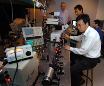
A Purdue-led research team developed a new method
to detect cancer by scanning surface veins with a laser. Ji-Xin
Cheng (foreground), an assistant professor of chemistry and
biomedical engineering, and graduate student Wei He, work with the
two-photon fluorescence microscope. The microscope is used to
detect circulating tumor cells labeled with tumor-specific
fluorescent probes developed by Philip Low (left), Purdue's Ralph
C. Corley Distinguished Professor of Chemistry. Postdoctoral
researcher Haifeng Wang (right) records the data displayed on the
computer screen. Low, Cheng, He and Wang co-authored a paper
published in the Proceedings of the National Academy of Sciences
detailing the cancer detection method and technology.
(Purdue News Service photo /
David Umberger)
|
Optical imaging provides high resolution and
chemical specificity for cancer detection, but it usually suffers from
limited penetration depth, making it hard to reach tumors inside the
body, said Ji-Xin Cheng, an assistant professor of chemistry and
biomedical engineering.
"In vivo detection of circulating tumor cells in surface veins
provides an excellent way to overcome this problem," Cheng said.
"Circulating tumor cells provide a benchmark for disease progression
and precise monitoring of their levels could lead to personalized
treatment," Low said. "This technique allows us to quantify the amount
of circulating tumor cells present, as opposed to tests that provide a
'positive' or 'negative' result.
"Through such precise monitoring, a physician could evaluate the
response to chemotherapy and regularly adjust the dosage so that only
the exact amount needed would be administered. This could reduce the
time a patient is treated and the serious side effects that occur."
The technique could provide doctors and patients results in a matter
of minutes and save the medical industry millions of dollars in
testing equipment, said Wei He, a graduate student in the Department
of Chemistry and the Department of Biomedical Engineering. He worked
on the project with Low and Cheng.
By directly labeling tumor cells while they are in the bloodstream,
some of the costs and problems associated with testing drawn blood
samples can be avoided, He said.
One sample can require five to 10 test tubes during the course of
sampling, processing and analysis such as handling, labeling and
washing," He said. "In addition, large hospitals can have more than
300 cancer patients in one day. Such a large influx can cause delays
in sample processing and delays can affect the results of analysis."
A paper detailing the technology and detection technique was published
in the July 10 Proceedings of the National Academy of Sciences. In
addition to Low, He and Cheng, postdoctoral researcher Haifeng Wang
and Lynn C. Hartmann, a professor of oncology and associate director
for education of the Mayo Clinic Cancer Center, co-authored the paper.
The technique uses a fluorescent tumor-specific probe that labels
tumor cells in circulation. When hit by a laser, which scans across
the diameter of the blood vessel 1,000 times per second, the tumor
cells glow and become visible. The in vivo flow detection was
performed on a two-photon fluorescence microscope in Cheng's lab. The
researchers compared several methods and found two-photon fluorescence
provides the best signal to background ratio. The technology is able
to scan every cell that is pumped through the vessel, He said.
Low's team has developed two labeling agents that attach to different
forms of cancer. One label targets ovarian, non-small lung, kidney and
endometrial cancer, and the other targets prostate cancer.
These labels would be administered through an injection. The first
label has already been tested in humans and has no adverse side
effects and could potentially be administered weekly, He said.
Computed tomography, or CT, scans and magnetic resonance imaging, or
MRI, are the current methods used to track the spread of cancer. These
methods have a limited resolution, and a 1 millimeter tumor could go
undetected by CT or MRI. The Purdue-developed technology can achieve
single-cell resolution and can detect rare cell populations.
"Our method can detect cancer cells early in disease development and
the test can be conducted frequently," Low said. "Discovering the
cancer early and knowing whether it has metastasized, or spread,
greatly improves a patient's chance for successful treatment."
The laser penetrates to a depth of 100 microns and is able to examine
shallow blood vessels near the surface of the skin. Advanced optical
technology could be incorporated into the technology platform and
enable the method to reach deeper vessels that handle larger volumes
of blood, Cheng said.
The Purdue team continues to work with the Mayo Clinic and is planning
to initiate a clinical trial to further evaluate the technique. The
team also plans to develop labels for additional types of cancer and
to downsize the equipment to make the technology portable.
This research was funded by an Indiana Elks Charities Grant, the
Purdue Cancer Center and an Ovar'Coming Cancer Together research grant.
|